
Digital radiography replaces traditional film with electronic sensors that record X‑ray data and convert it into instant images. Instead of waiting for chemical development, the sensor transmits the image directly to a computer where it appears on a monitor within seconds. This streamlined capture process allows clinicians to confirm image quality in real time and, when necessary, retake shots immediately to ensure diagnostic clarity.
There are a few common sensor types used in modern dental offices—direct sensors that send data immediately to the computer and indirect systems that scan a reusable plate. Regardless of the specific hardware, the end result is the same: a high-resolution digital file that becomes part of a patient’s electronic record. That digital file can be magnified, adjusted for contrast, and archived without the physical handling that film requires.
Because images are saved electronically, they integrate smoothly with practice management software and imaging suites. This integration helps clinicians track changes over time, compare current films with prior visits, and maintain a consistent visual record that supports accurate treatment planning and follow-up care.
One of the most tangible advantages patients experience is speed. Digital images appear almost instantly, shortening appointment times and reducing the need for repeat visits to obtain diagnostic images. The quick turnaround also makes it easier for dentists to discuss findings with patients during the same visit, improving understanding and helping patients make informed decisions about their oral health.
Radiation exposure from digital radiography is substantially lower than with conventional film X‑rays. That reduction results from more efficient sensors and the ability to capture quality images with less radiation. For patients who require frequent imaging—such as those undergoing orthodontic treatment or monitoring periodontal health—this reduction can make routine imaging safer over the long term.
Digital images are easily shared with specialists or other providers when coordinated care is necessary. Whether seeking a consultation with an endodontist, an oral surgeon, or an orthodontist, clinicians can transmit files electronically and receive feedback quickly, which helps streamline referral workflows and continuity of care.
Digital files can be manipulated with imaging software to highlight subtle findings that might be harder to see on film. Adjustments to brightness, contrast, and sharpness—or tools that allow magnification and measurement—help clinicians detect early decay, assess bone levels around teeth, and evaluate the integrity of existing restorations. These enhancement tools do not replace clinical judgment, but they extend the dentist’s ability to identify conditions at an earlier stage.
For procedures such as root canal therapy or implant planning, precise visual information matters. Digital radiographs provide clear views of root anatomy, canal morphology, and the relationship between teeth and surrounding structures. When combined with other diagnostic tools, these images contribute to more predictable treatment outcomes and more targeted clinical decision-making.
Because images can be compared side by side on the screen, clinicians can monitor disease progression or healing over time with greater accuracy. This longitudinal perspective supports preventive strategies and helps patients understand why certain treatments are recommended at specific points in their care.
From a safety standpoint, digital radiography’s lower radiation dose is a meaningful improvement for routine dental imaging. Modern sensors capture the necessary diagnostic detail more efficiently than film, allowing clinicians to achieve diagnostic-quality images with less exposure. The practice also follows established infection-control protocols for handling sensors and positioning devices to protect patient and staff safety.
Comfort can improve as well. Many contemporary sensors are thinner and designed to be less intrusive inside the mouth than older film packs, making the experience more tolerable for patients with sensitive gag reflexes or limited mouth opening. Clinicians take care to position sensors gently and communicate steps throughout the process to make each exposure as comfortable as possible.
There are environmental benefits too: digital imaging eliminates the chemical developers and fixers that film processing requires, reducing hazardous waste and chemical handling. Less physical storage space is needed since images are archived digitally, which simplifies recordkeeping and reduces the resources associated with maintaining paper and film files.
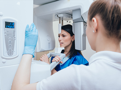
When you arrive for imaging, the dental team will explain the process and prepare you for a few short exposures. A small sensor or plate will be placed in the mouth in the area to be imaged; the dentist or hygienist will position the X‑ray arm outside the mouth to capture the image. The capture itself takes only a fraction of a second, and patients typically feel only brief pressure from the sensor while it is held in place.
After each exposure, the image is reviewed immediately on a monitor. If the team needs a clearer view, they can retake the image right away rather than scheduling a separate visit. The clinician will then review the images with you, pointing out any areas of concern and explaining how the findings relate to your overall treatment goals and preventive care plan.
Because images are added to your electronic chart, they become part of a long-term record that helps guide future care. Whether monitoring restorative work, tracking periodontal changes, or planning more complex treatment, digital radiographs give both patient and clinician a reliable visual reference to support shared decision-making.
At Comprehensive Family Dentistry, we use digital radiography to deliver precise, efficient, and patient-centered care. If you’d like to learn more about how digital imaging fits into your treatment or how we use these tools to support diagnosis and prevention, please contact us for more information.


Digital radiography uses electronic sensors to capture X-ray data and convert it into images that appear on a computer within seconds. Unlike traditional film, there is no chemical development process, which speeds diagnosis and reduces handling of physical materials. The instant display lets clinicians verify image quality immediately and retake exposures if needed to ensure diagnostic clarity.
Modern systems include direct sensors that transmit data instantly and indirect plates that are scanned after exposure, but both produce high-resolution digital files. Those files can be magnified, adjusted for contrast, and integrated into a patient’s electronic record for long-term tracking. Digital capture also simplifies comparison with prior images and supports more precise treatment planning.
Digital radiography typically requires much lower radiation doses than conventional film X-rays because sensors are more sensitive and capture usable images faster. Dental practices follow the ALARA principle—keeping exposure As Low As Reasonably Achievable—while using modern equipment and shielding when appropriate. These precautions make routine dental imaging safe for most patients when performed according to professional guidelines.
The practice also follows infection-control protocols for sensor handling and positioning to protect both patients and staff. If you have specific health concerns, such as pregnancy or recent medical imaging, discuss them with your dentist so exposures can be minimized or deferred if clinically appropriate. Your dentist will balance diagnostic needs with safety considerations to determine the best approach.
Before imaging begins, the dental team will explain the process and position a small sensor or plate in the area to be imaged. The X-ray arm is placed outside the mouth and the exposure itself takes only a fraction of a second, while patients generally feel only brief pressure from the sensor. Immediately after capture, the image is displayed on a monitor so the clinician can confirm quality and retake any shot if necessary.
Because images appear instantly, your dentist can review findings with you during the same visit and point out areas of concern using on-screen tools. The images are then added to your electronic chart to help guide treatment and future monitoring. Staff will take steps to maximize comfort and explain each step for a calmer experience.
Digital images can be enhanced with software tools that adjust brightness, contrast, and sharpness or provide magnification, which helps clinicians detect subtle signs of decay, bone loss, and restoration issues. Measurement and annotation features allow for precise assessment of root anatomy, canal morphology, and bone levels that are important for procedures like root canal therapy and implant planning. These capabilities extend the clinician’s ability to identify problems earlier and with greater confidence.
Side-by-side comparisons of current and prior images make it easier to monitor disease progression or healing over time and to evaluate the effectiveness of treatment. When combined with a clinical exam, enhanced imaging supports more targeted recommendations and improves predictability of outcomes. Clear visual information also helps patients understand proposed treatments and expected results.
Many contemporary digital sensors are thinner and designed to be less intrusive than older film packs, which can reduce discomfort for patients with sensitive gag reflexes or limited mouth opening. The smaller profile and quicker capture time often make the experience quicker and more tolerable for most people. Dental teams take care to position sensors gently and use patient-centered techniques to minimize pressure.
Some patients may still feel brief discomfort from holding a sensor in place, but staff will communicate throughout the process and make accommodations when possible. When intraoral imaging is not feasible or additional context is needed, clinicians may use extraoral imaging options to obtain the necessary diagnostic information. Comfort and diagnostic needs are balanced to ensure a safe, effective exam.
Digital radiographs are saved as electronic files and integrated into the patient’s digital chart, making them easy to archive, retrieve, and compare over time. These records are typically managed by practice software that creates secure backups and maintains a consistent visual history to support treatment planning and follow-up care. Electronic storage also eliminates the need for physical film storage and reduces the risk of lost or damaged records.
To protect patient privacy, images are maintained in secure systems with controlled access and routinely backed up to prevent data loss. When images must be shared with other providers, transmissions are encrypted and handled through secure channels in accordance with privacy standards. Patients can discuss record-handling practices with the office if they have questions about confidentiality.
Yes, digital radiographs can be transmitted electronically to specialists, labs, or other members of a treatment team, which speeds consultation and supports coordinated care. Files can be sent securely in common formats that preserve image quality and allow the receiving clinician to review and measure findings. Quick sharing reduces delays in referrals and helps ensure that all providers are working from the same diagnostic information.
Before images are released, the practice follows patient-consent and privacy procedures to ensure records are shared appropriately. Electronic sharing often results in faster feedback and can simplify collaborative treatment planning for procedures such as implants, endodontics, or orthodontics. Patients are encouraged to ask how their records will be used and who will have access.
The frequency of dental X-rays varies based on individual risk factors, oral health status, and treatment needs rather than a one-size-fits-all schedule. Patients with a history of decay, active periodontal disease, or ongoing orthodontic care may require imaging more often, while those with low risk and stable records may need fewer exposures. Your dentist will consider your medical and dental history, current exam findings, and professional guidelines when recommending imaging intervals.
Professional organizations provide reference guidelines, but the final decision rests on clinical judgment tailored to each patient. Regular visits and risk assessments help determine whether new images are necessary to diagnose changes or monitor treatment. If you have concerns about the timing of X-rays, discuss them with your provider to develop a plan that balances diagnostic benefit and safety.
Digital radiographs are particularly effective at revealing interproximal decay (decay between teeth), early bone loss from periodontal disease, hidden infections or abscesses at tooth roots, and the position of unerupted or impacted teeth. They can also show the condition of existing restorations and help identify fractures or other internal tooth problems that may not be visible during a clinical exam. Early detection allows clinicians to recommend less invasive treatments when appropriate.
For surgical or restorative planning, digital images clarify root structure and the relationship between teeth and supporting bone, which improves case selection and execution. While radiographs are a powerful diagnostic tool, they complement rather than replace a thorough clinical examination. Combining visual, tactile, and radiographic information leads to the most accurate diagnosis and effective care.
At Comprehensive Family Dentistry, digital radiography is used to deliver precise, efficient, and patient-centered diagnostic information that supports individualized treatment planning. Immediate image review allows the dentist to explain findings during the same visit and use on-screen tools to show patients what is happening in their mouths. Electronic records make it easy to compare past and present images, which helps track healing and disease progression over time.
The practice emphasizes safety, comfort, and clear communication when using digital imaging, and staff work to minimize exposure while maximizing diagnostic value. Digital files also facilitate timely collaboration with specialists when multidisciplinary care is needed, helping to coordinate treatment and improve outcomes. Patients are encouraged to ask questions about their images so they can participate in decisions about their oral health.

Whether you’re ready to schedule your next dental appointment or simply have questions about our services, connecting with our team has never been easier.
Our friendly team is here to assist with appointment scheduling, answer questions about treatments, and address any concerns you may have. You can call or use our convenient online form to get in touch.
Take the first step toward a healthier, more confident smile—contact us today and experience the difference personalized dental care can make.

Back to top